
Indications
When to Call Mobile Pet Imaging
Our top requests are for head, chest, and abdomen high definition CT Scans, but there are hundreds of reasons why your pet would need a CT.
Head
Click text to see more
Any Mass or Swelling in the Head (eyes, jaw, ears, nose, etc.)
- CT is an excellent diagnostic tool for any swelling or possible mass in the head. It will help identify any skull abnormalities which may be causing decreased appetite, lethargy, pain, physical discomfort, etc.
Inner vs Middle vs Outer or External Ear Disease (Bulla)
- CT can evaluate even the smallest of changes in the mouth. A scan will help find a cause for your pet having a decreased appetite, nasal discharge, drooling excessively, odor coming from the mouth, and/or being uncomfortable while eating.
Dental Disease
- CT can evaluate and distinguish between inner, middle, and outer/external ear canal disease. This is very important if your pet has a head tilt, hearing loss, walks in circles, and/or balance problems (vertigo).
Upper Respiratory Sounds (stridor, snoring)
- CT can help evaluate causes for upper respiratory sounds. Anything which may cause an obstruction in airflow may cause stridor, snoring, or reverse sneezing. Airflow is usually disrupted by a blockage or collapse in the larynx (voice box) or trachea (windpipe). This may make your pet seem out of breath (panting) at times as well as being very uncomfortable. Common conditions diagnosed with CT are masses in the pharynx, larynx, trachea, enlarged lymph nodes or foreign bodies.
Chronic Ear Infections (Chronic Otitis)
- CT can help identify causes for your pet’s persistent ear canal infection. Chronic, long lasting or recurring ear infections have many causes such as resistant bacteria and/or yeast or fungus, allergies, growths, and skin parasites. These ear infections typically cause itchy, painful, sometimes swollen (ear hematoma), and often smelly ears. If the main cause of the ear infection is not treated, and it continues to happen, it may lead to rupturing of the eardrum (tympanic membrane) and/or narrowing of the ear canal. CT can identify masses, fluid buildup and foreign bodies which could be causing the ear disease.
Salivary Gland/Lymph Node Evaluation
- CT can help identify causes for swelling under and around your pet’s jaws. Some common causes for the swelling are the lymph nodes or salivary glands. When a salivary gland is infected it is called sialadenitis. Salivary glands can also become obstructed. These conditions, which can be very painful, can be caused by bacteria, inflammation, systemic diseases, trauma from penetrating wounds, or a stone blocking the duct. Other possible causes for swelling under the jaws are benign (non-cancerous) masses vs malignant (cancerous) masses. Lymphoma which is a type of cancer that affects the lymph nodes is one of the most common causes. Lymph nodes may also be reactive due to an infection caused by dental disease.
Growths in the Back of the Nose, Throat, and Mouth (Nasopharyngeal polyp)
- Nasopharyngeal polyps are common, benign (non-cancerous) growths that are found at all stages of life but more commonly seen in kittens. These polyps are typically caused by chronic inflammation and swelling due to upper respiratory infections. Feline respiratory viruses such as calicivirus or herpesvirus are typically at fault. A CT scan can help diagnose these growths since it will allow complete evaluation of all the structures in the head and thereby help your veterinarian prepare for surgical removal.
Head Trauma or Injury
- After an unexpected event, such as being hit by a car or an acute hit to the face, a CT scan can identify the full extent of damage to determine the best course of action for your pet. CT is a great tool that gives your veterinarian 3D images of your pet. If surgery is needed, Mobile Pet Imaging can provide your veterinarian with 3D images and can plan the surgery before your pet is anesthetized.
Pituitary Gland Tumor
- If your pet begins drinking excessive amounts of water, urinating more than normal, has an increased appetite, pants excessively, and appears to have a “pot-bellied” abdomen, a CT scan could be very helpful. A possible cause for this abnormal and unusual behavior is Cushing’s disease or hyperadrenocorticism. A CT scan can help differentiate between adrenal gland and pituitary dependent Cushing’s disease. Macroadenoma (a large non-cancerous tumor of the pituitary gland) can also be diagnosed and the results of the CT scan can help determine the outcome (prognosis) and what treatment strategy would be more beneficial for your pet.
Tonsils Evaluation
- Inflammation of the tonsils or tonsillitis may occur in dogs causing difficulty eating or swallowing, drooling, discomfort, lethargy, and others. A CT scan can help determine possible causes for tonsil disease and will provide further information for treatment.
Nasal & Sinus
(Click text to see more)
Runny Nose/Nasal Drip/Nasal Congestion (Chronic Nasal Discharge)
- If your pet seems like it has the common cold, an infection may be the root cause of the nasal discharge. Nasal mucus or pus may be an indication of a viral, bacterial, or fungal infection. Autoimmune diseases or dental disease could also be the cause of the persistent nasal discharge. A CT scan may help differentiate the cause and will show if there is destruction of the nasal passages. A CT is a great option if regular antibiotic and viral therapy has not shown resolution of the nasal discharge.
Swelling of the nose (Nasal Swelling)
- Many reasons could lead to the swelling of the nose. A CT scan could help differentiate the leading causes such as bony abnormalities, tumors, injury, foreign object, etc.
Nose Bleed (Epixtasis)
- Nose bleeding is something that could potentially be very serious and even possibly life threatening. It should be immediately investigated and treated. A CT scan is one of the best imaging tools to help identify a cause. Different causes such as benign trauma, foreign objects, immune mediated diseases, parasitic infections, and even cancer could be the cause for nosebleeds.
Nasal Deformity
- CT may help identify nasal deformities, which may often cause a “runny” nose, nasal discharge, congestion, etc. These deformities may often be due to trauma but may also sometimes be a birth defect (congenital). Cats with rhinitis, nasal discharge, sneezing, and/or loud “congested” breathing may be due to a deformity of the nose. Some immune mediated conditions may also cause the nose to become deformed.
Pulmonary (Lungs) and Thorax (Chest)
Click text to see more
Mediastinal Disease, Mediastinal Mass, Air In the Chest (Pneumomediastinum)
- The mediastinum is the area of your pet’s chest between the lungs, from the sternum to the spine where the heart and other organs are found. Mediastinal disease is caused by trauma, infection, inflammation, growths, cancer, or foreign bodies. Many of these problems can be missed on x-rays but seen on a CT scan. If there is any suspicion for air, a mass, or anything suspicious in your pet’s chest radiographs, a CT scan is the best diagnostic tool to help resolve the suspicion.
Further Evaluation of Pulmonary Disease as Seen on X-rays
- If your pet has been diagnosed with cancer, typically the next step is to perform X-rays on his or her lungs to see if the cancer is spreading. A CT scan shows much more than an X-ray so the cancer would be diagnosed much earlier than with x-rays. CT scan will detect spreading a lot sooner than radiographs- in order for nodules to be detected by radiographs, they need to be 8-10X larger than with CT scans. If your pet has been recently diagnosed with cancer, a CT scan of the chest should be performed to determine your pet’s prognosis (expected outcome).
Masses in the Chest/Spreading of Cancer in the Chest (Pulmonary Metastasis)
- If your pet has been diagnosed with cancer, typically the next step is to perform X-rays on his or her lungs to see if the cancer is spreading. A CT scan shows much more than an X-ray so the cancer would be diagnosed much earlier than with x-rays. CT scan will detect spreading a lot sooner than radiographs- in order for nodules to be detected by radiographs, they need to be 8-10X larger than with CT scans. If your pet has been recently diagnosed with cancer, a CT scan of the chest should be performed to determine your pet’s prognosis (expected outcome).
Evaluating the Source of Air in the Chest (pneumothorax) or Air in the Mediastinum (membrane partition between the lungs) (pneumomediastinum)
- CT is a tool that is very helpful in identifying causes for air in the thorax, whether it be due to trauma, foreign body, neoplasia (cancer), etc. It can help identify the source and help with the next course of action. A common cause for pneumothorax is the puncturing of a lung due to a broken rib. Another common cause is a pulmonary bulla (dilated air space in the lung) which may rupture and leak air into the pleural space. CT can help pinpoint the cause of the air in the chest and, therefore, the treatment will be done quickly (no need for multiple other tests or radiographs) and the outcome improves in most cases.
Pulmonary Masses
- Chronic coughing that does not resolve with coughing medications or antibiotics could be due to a more serious problem. If no evidence of heart disease is noted, a CT scan is recommended. In some cases the radiographs don’t show masses that will be seen on a CT scan. In cases where a thoracic mass is noted on x-rays, a CT scan helps create a surgical plan and determine if other masses are present which were missed by the x-rays. A CT scan will also allow the surgeon to make sure surgery is even a possibility prior to surgery (ie, surgical planning). This is done by evaluating the vessels which are in the vicinity of the mass.
Mediastinal Disease or Masses
- These masses form in the area of your pet’s chest that separates the lungs. Although mediastinal disease is uncommon, some of the causes are cranial mediastinal lymphoma (a type of cancer), thymoma (tumors from the lining of the thymus gland), and ectopic thyroid carcinoma (another type of cancer in an abnormal place). One of the main reasons this is considered is due to persistent elevation in the calcium (hypercalcemia) blood levels. Therefore, if your pet has persistent elevated calcium blood levels, a CT of the thorax could help identify mediastinal disease if that is the cause. In those cases, the abdomen is also included to make sure no masses or spreading has occurred in the abdomen.
Pleural Disease (Membranes around the lungs) or Masses
- Some of the most common pleural diseases are pleural effusion (abnormal accumulation of fluid within the chest cavity), pneumothorax (accumulation of air in the space between the chest wall & lungs), chylothorax (accumulation of “milky fluid” within the chest cavity), hemothorax (accumulation of blood in the chest cavity), and pleural nodules/masses. A CT scan is an excellent tool to help identify the cause of these diseases.
Thoracic Wall Disease or Masses
- If your pet develops a growth (i.e. mass, swelling, etc.) or suffers any type of injury to and around the chest wall and/or underarm (axilla), a CT can help evaluate the entire area for a better understanding of the disease or abnormality. It also helps you and your veterinarian plan the best treatment possible. A CT scan will also help determine the extent of the growth or mass, which determines if it is resectable (able to be removed by surgery) or not. Mobile Pet Imaging may be able to provide your veterinarian 3D views if appropriate to aid in the planning of surgery.
Distinguish Lung vs Mediastinal or Pleural Masses
- A CT scan may help differentiate all the different diseases that may occur in the chest (thorax) and it is an excellent tool for surgical planning. Since a CT scan has 3D reconstruction, it will be able to differentiate better than x-rays and ultrasound in most cases.
Suspect Heart Base Masses
- If there is any suspicion for a heart base mass, a CT scan is an excellent tool to help diagnose this. Hemangiosarcoma is one of the most common form of heart tumor. Although rare, it is very aggressive. Typically patients suffering from this are first noted with weakness, lethargy, and sometimes difficulty breathing.
Suspect Thoracic Lymphadenopathy (enlarged lymph nodes in the chest)
- Lymph nodes are an integral part in the functioning of the dog’s immune system. So when there is an infection or some type of inflammation, they may become enlarged in size. Another common cause for enlarged lymph nodes is, unfortunately, cancer. The most common type of cancer which affects the lymph nodes is called lymphoma. A CT scan will be able to pick up enlarged lymph nodes much earlier than x-rays and may be able to improve the prognosis (expected outcome) if caught in time.
Suspect Heart BaEvaluating Chronic Pneumoniase Masses
- When evaluating cases with chronic pneumonia, CT is an advanced imaging tool that will be able to monitor improvement or worsening much better than radiographs (x-rays). Most common pneumonias are typically caused by a bacterial infection, although some fungal infections may also cause pneumonia. Aspiration pneumonia is also commonly seen and is due to a relatively large amount of material from the mouth, esophagus or stomach entering into the lungs after being “aspirated”. Finally foreign objects can cause pneumonia and CT is an ideal tool to diagnose them; most times they are missed by X-rays. If your veterinarian wants to perform a bronchoscopy and /or needle aspirates (a way to obtain a sample for diagnosis) of the lungs, a CT is the best method for planning those procedures.
Suspect Pulmonary Fibrosis
- Pulmonary Fibrosis is a condition that typically results in inflammation and scarring of the tiny air sacs of the lungs and lung tissue. When suspecting pulmonary fibrosis, a CT scan is an advanced imaging tool that may help diagnose as well as determine the severity of the condition. The more severe this disease becomes, the less oxygen is passed into your pet’s body tissues after every breath.
Spinal
Click text to see more
Slipped Disc or Intervertebral Disc Disease (IVDD) in Young Chondrodystrophic (cartilage maldevelopment) Patient
- A slipped disc can cause a variety of clinical signs including back pain, weakness (affecting one or multiple limbs) to even paralysis and the inability to walk or stand. Some of the breeds most commonly affected by this condition include the Dachshund, Pekingese, Shih Tzu, Corgi, and Bulldog. Depending on the clinical presentation a CT scan with or without a myelogram
Spinal (vertebral column or backbone) Masses
- CT can evaluate Spinal masses or tumors can occur in our pet friends and cause multiple symptoms such as pain, limping or even paralysis. CT can help in the diagnosis of this masses or tumors since iteven the smallest of changes in the mouth. A scan will help find a cause for your pet having a decreased appetite, nasal discharge, drooling excessively, odor coming from the mouth, and/or being uncomfortable while eating.
Lumbosacral Disease
- Lumbosacral disease affects the lower spine and may cause pain, walking difficulties, urinary incontinence or trouble defecating. CT scans evaluate the lumbosacral region and its surrounding structures and sometimes use dynamic studies to improve diagnostic accuracy.
Spinal Pain/Inflammation (discospondylitis)
- Discospondylitis is an infection‑related inflammatory disease of the spine leading to back or abdominal pain, hunched posture, fever or paralysis. CT imaging detects this condition earlier than X‑rays.
Spinal Fractures or Luxations (dislocations) due to Trauma
- CT scans evaluate spinal fractures or dislocations following trauma. Detailed 3D images help veterinarians plan treatment and determine whether surgery is necessary.
Muscles & Bones
Click text to see more
Front Legs (forelimbs or thoracic limbs): Metacarpal, Carpal, Elbow, and/or Shoulder Disease Affecting any Bone or Joint
- The diagnosis of forelimb lameness (limping) in pets can be very exhaustive and challenging for your veterinarian. Lameness of the elbow and/or shoulder most of the time requires many diagnostic methods. Elbow lameness in our canine patients can be caused by conditions such as joint incongruity, fragmented coronoid process (FCP), osteochondrosis of the medial humeral condyle and ununited anconeal process (UAP). These conditions are grouped under the term “Elbow Dysplasia”. One or multiple conditions can occurs at the same time. Some symptoms include limping, decreased range of motion, crying, and swollen joints. In many cases, performing a CT scan will yield valuable information in order to determine the best treatment, plan, and prognosis. However in some cases CT scanning would be used in addition to arthroscopy and/or MRI to further diagnose some of these conditions.
Shoulder Lameness
- Shoulder lameness can be caused by different conditions which include medial (closest to the midline) shoulder instability, osteochondritis dissecans (OCD - damaged cartilage), osteoarthritis (DJD - degenerative joint disease) and biceps tendinopathy (inflammation of the bicep tendon). These conditions can occur by themselves or multiple conditions can be noted in one patient. In many cases, performing a CT scan will yield valuable information in order to determine the best treatment plan and prognosis. However in some cases CT scanning will be used in addition to arthroscopy and/or MRI to further diagnose some of these conditions.
Back legs (hind limbs or pelvic limbs): Metatarsal, Tarsal, Stifle, and/or Hip Disease Affecting any Bone or Joint
- The diagnosis of hindlimb lameness (limping) in pets can be very exhaustive and challenging for your veterinarian. A CT scan can help identify many conditions affecting the pelvic limbs such as stifle (“knee”) or tarsal (hock or “ankle”) joint OCD (osteochondritis dissecans), hip disease (i.e. hip dysplasia, legg-calve-perthes disease), limb swellings and/or growths, bone tumors (cancer), myositis (inflammation of the muscle), abscess formation, etc.
Acute Trauma (Complicated Long Bone, Joint or Pelvic Fractures)
- Unfortunately, trauma can occur at any given time and affect our precious pet friends. If your pet suffers any trauma or is suspected to have experienced trauma, a CT scan will help evaluate the affected areas by providing a 3-dimensional picture of the region(s) of interest including all bones and soft tissue structure. This complete and accurate evaluation of the affected areas is essential in order to determine the best treatment plan possible.
Appendicular Skeleton Masses (Muscle, Cutaneous, Subcutaneous and/or Bone)Ear Infections (Chronic Otitis)
- The appendicular skeleton consist of the bones of the forelimbs and hindlimbs. Masses can appear in the muscles, on the skin (cutaneous), under the skin (subcutaneous), and in or on the bones. Did you know dogs have approximately 100 more bones than humans and cats have approximately 44 more bones than humans? A CT scan will help evaluate the organ of origin and determine the extent of the mass. This information will be valuable when determining the best treatment plan and will also aid with the prognosis.
Unexplained Lameness (limping or gait abnormality)
- The diagnosis of an unexplained lameness in pets can be very costly, frustrating, exhaustive, and challenging. Performing a CT scan of the suspected limb(s) and/or spine is a quick, cost-effective, and excellent diagnostic that will yield valuable information in order to reach the diagnosis faster and determine the best treatment plan and prognosis. However, in some
Shoulder, Stifle (“knee”) or Tarsal (“ankle”) joint OCD in the Young Patient
- Shoulder lameness can be caused by different conditions which include medial (closest to the midline) shoulder instability, osteochondritis dissecans (OCD - damaged cartilage), osteoarthritis (DJD - degenerative joint disease) and biceps tendinopathy (inflammation of the bicep tendon). These conditions can occur by themselves or multiple conditions can be noted in one patient.
- Similarly, conditions affecting the pelvic limbs such as stifle (“knee”) or tarsal (hock or “ankle”) joint OCD (osteochondritis dissecans) can affect our pets even at a young age. In many of these cases, performing a CT scan will quickly yield valuable information in order to determine the best treatment plan and prognosis. However in some cases CT scanning will be used in addition to arthroscopy and/or MRI to further diagnose some of these conditions once localized to a specific bone or joint.
Spinal Fractures
- Unfortunately, spinal fractures can occur at any given time and affect our precious pets. Your pet may have been hit by a car, attacked by another animal, or fallen from a considerable height. Symptoms may include spinal pain and the inability or reluctance to walk or stand. Performing a CT scan can help diagnose these fractures and assist your veterinarian in the proper treatment or surgery for your pet. In some cases CT scanning will be used in conjunction with MRI.
Skull Fractures
- Skull fractures typically occur after some sort of trauma (i.e. hit by a car, attacked by another animal, fallen from a considerable height, etc.). These fractures can be very serious and have many symptoms including head tilt, circling, blindness, seizures, other neurological symptoms, and coma amongst others. A CT scan can help identify these fractures and assist your veterinarian in the proper treatment or surgery if necessary. In some cases CT scanning will be used in addition to MRI.
Pelvic
Click text to see more
To evaluate intrapelvic masses and lymph nodes
- A CT scan is an excellent diagnostic imaging modality to evaluate the pelvis which is made up of the following bones: ilium, ischium, and pubis. The front part of the pelvis is attached to the sacrum (part of the spine) via the sacroiliac joint.
- The CT will allow complete evaluation of all the intrapelvic structures (see below) in order to determine the best course of action.
Abdomen
Click text to see more
Hepatic masses (Liver Masses)
- Evaluation of any liver masses, to see if they are surgically resectable, is another reason to perform a CT scan of the liver. CT can help evaluate the vascularity (blood supply) of liver (hepatic) masses which is particularly important if tumor embolization (shutdown or decrease blood supply) is desired prior to surgery.
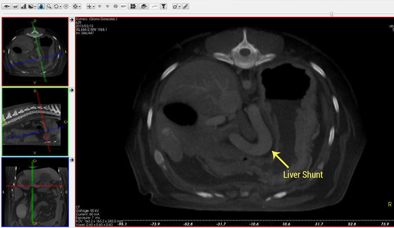
Liver Shunt – Congenital or Acquired Condition
- A portosystemic shunt (PSS), also known as a liver shunt, is not necessarily that common in dogs, but in certain breeds (Yorkies, Jack Russell Terriers, Cocker Spaniels, Lhasa Apsos, Poodles, Labs) they are more common. If your dog develops liver disease it may have a liver shunt. If your puppy begins acting strangely (staring into space, acting disoriented, circling or head pressing, having seizures) a liver shunt is something that needs to be investigated and potentially may be surgically corrected. These patients may or may not have elevated liver enzymes but almost always have an elevated bile acids test. If your pet has elevated bile acids, a CT scan is the ideal imaging tool to evaluate the liver and identify any possible liver shunts (abnormal vessel). CT scans are superior to an ultrasound when evaluating for liver shunts.
Surgical Planning
- For most kinds of abdominal surgery it is always best to have a CT scan before the procedure. This gives your veterinarian a 3D view before the surgery and can help him or her plan appropriately and this will help improve the outcome of the surgery.
Abdominal Mass (liver, spleen, kidneys, adrenal glands, stomach, prostate, intestines, lymph node, etc.)
- CT is ideal for evaluating any suspicion of a possible abdominal mass. A CT scan will not only be able to identify if there is a mass but it will also determine the location and provide information for surgical planning. CT will also be able to identify any possible evidence of spreading of metastatic neoplasia (cancer). One of the most common CT scans we do at Mobile Pet Imaging are that of adrenal masses- the CT helps plan the surgery and, therefore, helps improve the outcome of such a challenging surgery.
Enlarged Abdominal Organs (Organomegaly)
- A CT scan can help evaluate your pet’s intra-abdominal organs in case any of them become enlarged. Intra-abdominal organs such as the liver, spleen, pancreas, kidneys, etc. can become enlarged secondary to trauma, infection, cancer, immune-mediated conditions, etc.
Ectopic Ureter, Ureteral Disease (IVP/IVU)
- CT can help identify causes for swelling under and around your pet’s jaws. SomeIf your puppy has many episodes of urinating everywhere or is constantly leaking urine a CT scan will help diagnose this problem. This problem occurs because the ureter (tiny tube) which carries the urine from the kidney to the bladder is not connected properly. This disease is caused by abnormal development in the embryo. Since the urine doesn’t pass through the urinary bladder there is nowhere to store the urine. Therefore your pet is constantly “urinating” or dribbling urine. CT is the imaging modality of choice to help diagnose this problem and create a plan of action for surgery. common causes for the swelling are the lymph nodes or salivary glands. When a salivary gland is infected it is called sialadenitis. Salivary glands can also become obstructed. These conditions, which can be very painful, can be caused by bacteria, inflammation, systemic diseases, trauma from penetrating wounds, or a stone blocking the duct. Other possible causes for swelling under the jaws are benign (non-cancerous) masses vs malignant (cancerous) masses. Lymphoma which is a type of cancer that affects the lymph nodes is one of the most common causes. Lymph nodes may also be reactive due to an infection caused by dental disease.
Diaphragmatic Hernia
- The diaphragm is a muscle between your pet’s chest and abdomen that is involved during breathing. A diaphragmatic hernia is a defect or tear in this muscle which can occur as a birth defect (congenital) or after trauma. This defect can cause breathing abnormalities due to abdominal organs moving into the chest and occupying space where the lungs should be. A CT scan can provide valuable information regarding the size and nature of the hernia as well as what organs may be moving through the defect at any given time. The CT helps with the treatment and planning for a course of action which in most cases will include surgery to fix the defect.
To Evaluate Organ of Origin, Invasion, Resectability and Vascularity of Abdominal Masses
- CT is ideal for evaluating abdominal masses. A CT scan will not only help identify the organ of origin but it will also determine the location and provide information for surgical planning (i.e. size, vascularity, presence of adhesions, resectability potential, etc.). CT will also be able to identify any possible evidence of spreading of neoplasia (cancer).
Caval Invasion with Hepatic and Adrenal Tumors
- A CT scan can help evaluate the vena cava (large vein) or aorta for tumor invasion in cases of liver or adrenal gland tumors. The liver and the adrenal glands live in close proximity to the vena cava and therefore when they develop tumors, these can grow into the nearby vena cava. CT angiography allows for the evaluation of the inside, or lumen, of blood vessels and organs of the body, with special interest in the veins and arteries. This is done or achieved by the intravenous administration of a contrast agent prior to and/or during the CT scanning process.
Benign or Metastatic Hepatic Lesions
- When liver lesions are difficult to interpret via X-rays and/or ultrasound, a CT scan will give further information and it may even help distinguish between malignant (cancerous) and benign (not cancerous) lesions of the liver. It also helps your veterinarian determine if surgery is needed.
To Differentiate Between Peritoneal or Retroperitoneal Masses or Lesions
- Depending on their anatomical location, the organs in the abdominal cavity can be described as peritoneal or retroperitoneal. Anything that affects these organs can cause abnormalities which can make the distinction between organ location difficult. A CT scan can help clarify the location of the organ or lesion in question so that the best treatment plan can be pursued.
Pancreatic Tumors/Masses or Insulinoma
- Insulinomas are tumors that arise from the pancreas. These tumors can cause a drop in your pet’s blood sugar levels which can cause fainting, weakness, collapse, seizures, etc. A CT scan can help find abnormal tissue and/or masses on the pancreas which will ultimately allow planning for the best treatment (i.e. surgery vs other). A CT scan is superior to an ultrasound when evaluating for insulinoma or other pancreatic tumor.
To Evaluate for Chronic GI Loss (small GI masses)
- CT can help diagnose gastrointestinal disease by providing a non-invasive survey evaluation of the entire tract (including the stomach, small intestine, colon, etc.) in one procedure. Sometimes small masses can be missed on X-rays or ultrasound due to their size and/or location but they can be diagnosed via CT.
Q: To Evaluate and Clearly Define Abdominal Wall Masses or Swelling
- CT is ideal for evaluating abdominal wall masses or swelling. A CT scan will not only help identify the organ of origin but it will also determine the specific location and provide information for surgical planning (i.e. size, vascularity, presence of adhesions, resectability potential, etc.). CT will also be able to identify any possible evidence of spreading of metastatic neoplasia (cancer).
Clot Formation – Aortic, Venous, and/or Portal Thrombus Formation, Tumor Thrombus, etc.
- CT angiography allows for the evaluation of the inside, or lumen, of blood vessels, which can be very useful when looking for thrombus (blood clot impeding blood flow) or clot formation. This is done or achieved by the intravenous administration of a contrast agent prior to and/or during the CT scanning process. Clots can form in any vessel but when they occur, they are typically seen in the major vessels such as the aorta, vena cava or portal vein. Embolus (when a piece of the clot breaks off and travels through the vessels until it reaches a vessel too small to let it pass), can also be identified with CT angiography.
GI Foreign Bodies
- A recent study showed that CT scans are as good as ultrasounds when looking for foreign bodies in the intestines. Traditionally radiographs and ultrasound are the modalities that are used as a first options, however in cases when ultrasound may not be available, a CT is a proven choice.
Mouth & Neck
Click text to see more
Dental Disease
- CT can evaluate even the smallest of changes in the mouth. A scan will help find a cause for your pet having a decreased appetite, nasal discharge, drooling excessively, odor coming from the mouth, and/or being uncomfortable while eating.
Neck Swelling (Salivary Gland / Lymph Node Evaluation)
- CT can help identify causes for swelling under and around your pet’s neck. Some common causes for the swelling are the lymph nodes or salivary glands. When the salivary gland is obstructed it is called sialandenitis. This condition, which can be very painful, can be caused by infection, inflammation, systemic diseases, trauma from penetrating wounds, or a stone blocking the duct. Other possible causes for swelling under the neck are benign (non-cancerous) masses vs malignant (cancerous) masses. Lymphoma which is a type of cancer that affects the lymph nodes is one of the most common causes. Lymph nodes may also be reactive due to an infection caused by dental disease.
Neck Masses
- If your pet develops a neck swelling or lump it may be due to a mass and a CT scan is an excellent diagnostic tool to evaluate the tissue of origin (lymph nodes, thyroid glands, esophagus, trachea, jugular veins, carotid arteries, cervical spine, muscles or subcutaneous tissues), invasion into adjacent structures, resectability and vascularity. The CT will provide vital information for the best treatment plan and prognosis.
General
Click text to see more
Pre‑Surgical Evaluation of Any Tumor
- Pre-Surgical Evaluation of any tumor, either soft tissue or osseous (ossified or turned into bone), and staging of the disease or condition. Staging is where the rest of the body is checked to make sure a tumor has not spread. A CT scan will provide valuable information as far as the tissue of origin, invasion into adjacent structures, resectability potential and vascularity. In addition to scanning the region of interest, oftentimes staging of the disease is achieved by performing a CT scan of the thorax to evaluate for potential spreading (metastasis) when cancer is suspected or diagnosed.
Pre‑Surgical Evaluation of Angular Limb Deformities (curved or bowed limb)
- Limb deformities are either congenital (from birth) or developmental. Puppies and kittens do most of their growing between 4-8 months. Growth plates are at the ends of the long bones and are the softest part of the bones. Therefore if your pet is injured (falls from a significant height, hit by a car, etc) the growth plates can close and cause the bones to stop growing or become twisted/deformed. CT scans are instrumental and more superior to radiographs in establishing a diagnosis and planning a treatment strategy. 3D reconstruction of the affected bone(s) by our computers aid tremendously in planning a treatment for the patient.
CTA - Computed Tomography Angiography
Angiography refers to the evaluation of the inside, or lumen, of blood vessels and organs of the body, with special interest in the veins and arteries. This is done or achieved by the intravenous administration of a contrast agent prior to and/or during the CT scanning process.
One of the most common indications for which we perform abdominal CT scans is to evaluate for the presence of a portosystemic shunt (PSS) or liver shunt- such as the one shown here.