
CT Scans
Case Studies
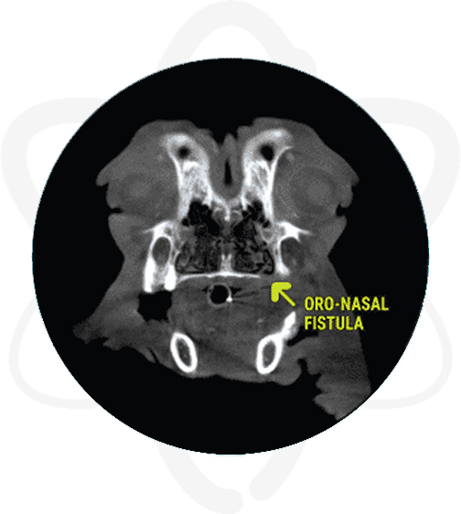
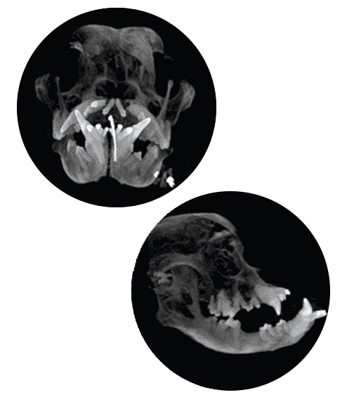
10 Year Old English Bulldog with Nasal Discharge

Overview
Signalment and History
Patient is a 10 yr old, FS, English Bulldog with unresolved nasal discharge. CT scan of the skull was requested for further evaluation since dental radiographs and cleaning did not reveal a cause for the discharge


Diagnostics Prior to CT Scan
- CBC/Chem/UA/T4: unremarkable
- Intra-oral radiographs- no cause for the nasal discharge noted
Pertinent Findings From Our Board-Certified Radiologist's Report
- Oro-nasal fistula left palatine bone level with triadan 207, the defect is completely covered by gingiva
- Periodontal disease 311 & 411 and multiple absent teeth
- Incidental dystrophic mineralization of both external ear canals
How CT Helped in This Case
- Allowed complete dental, nasal and sinus evaluation in one procedure.
- Discovered oro-nasal fistula not appreciated on intraoral radiographs (IOR) which can explain the clinical signs and provided detailed information for surgical planning.
Benefits of CT in Dental Cases
In cases of dental disease, dental X-rays are a great modality as an initial step especially when evaluating specific details of the teeth (i.e. types of resorption, periodontal ligament, lamina dura cases, etc). However, in unresolved cases in which IOR (intra-oral radiographs) have not yielded a cause, CT becomes an important tool since it can reveal lesions that superimposition would make impossible to see on IOR, such as oronasal fistulas. CTs are also useful in cases of maxillofacial fractures, tumors/cysts and TMJ pathology. As technology advances, CT scans will play a more important role in the evaluation of dental and nasal structures.
A 2016 study states “it is unnecessary to routinely perform IOR following CT (when acquired and reconstructed in 0.5 or 1 mm slice thickness) when testing for endodontic disease in dogs because there is a high level of agreement between techniques and between observers. This recommendation also is true for periodontal disease, except for when evaluating the mandibular incisors.“1 At MPI, our slices are at least 0.6mm, meeting this requirement. Our equipment performs a dental CT scan in only 30 seconds, which makes anesthesia time very short (10–20 mins. pre and post contrast).
1 Campbell RD, Peralta S, Fiani N and Scrivani PV (2016) Comparing Intraoral Radiography and Computed Tomography for Detecting Radiographic Signs of Periodontitis and Endodontic Disease in Dogs: An Agreement Study. Front. Vet. Sci. 3:68.