
CT Scans
Case Studies
6 Month Old Kitten With Abnormal Behavior


Overview
Signalment and History
Patient is a 6 month old, female, DSH who was referred for CT angiography (portogram) to determine if a liver shunt was present due to elevated bile acid profile. Patient had copper colored irises


Diagnostics Prior to CT Angiography
- CBC, chemistry and urinalysis- unremarkable
- Bile acid profile- preprandial- 206.2, postprandial- 208.9 (normal postprandial <20)
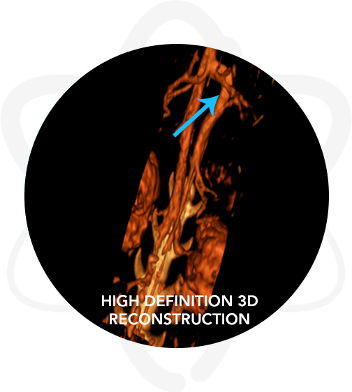
High-Definition CT Angiography
Patient was anesthetized per our anesthesiologist’s recommendations. A pre-contrast CT scan of the abdomen was performed. Afterwards, Omnipaque™ (contrast agent) was administered IV and post-contrast studies (CT angiography) were acquired.

Pertinent Findings From Our Board-Certified Radiologists's Report
- Single extrahepatic portosystemic shunt (splenophrenic)
- Bilateral renomegaly – likely secondary to the shunt
Following the CT Angiography
Patient did well on medical management until surgical correction of the abnormal liver blood-vessel was performed.