
CT Scans
Case Studies
11 Year Old German Shepherd With History of Coughing


Overview
Signalment and History
Patient is a 11 yr old, FS, German Shepherd Mix with a 4-month history of coughing that improves while she is on antibiotics.


Diagnostics Prior to CT Scan
- Chemistry panel: Ca 12.1
- Radiographs: Broncho-interstitial pattern
- Tracheal wash: epithelial cell hyperplasia with suppurative and septic inflammation.
- Tracheal culture: Resistant E. Coli
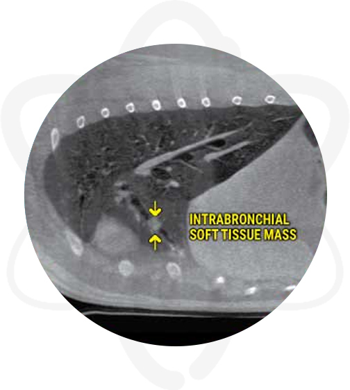
High-Definition CT Scan of the Thorax
Patient was anesthetized and a pre contrast CT scan of the thorax was performed. Afterwards, Omnipaque ™ (contrast agent) was administered IV and a post-contrast study of the thorax was acquired.
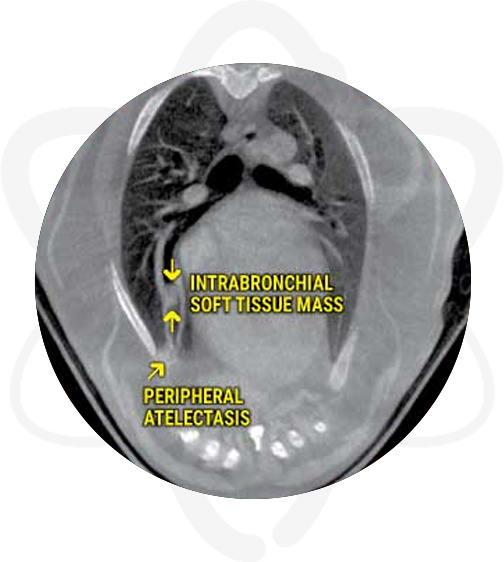
Pertinent Findings From Our Board-Certified Radiologist's Report
- Intrabronchial soft tissue mass lesion right middle lung lobe with complete obstruction
- Mild resorption atelectasis of the ventral peripheral aspect of the right middle lung lobe
- No evidence of metastasis in other lung fields and thoracic structures (or intrathoracic structures)
- Spondylosis deformans
How CT Helped in This Case
CT found a mass that was missed on radiographs which explain the history of the cough and made a difference in the outcome by providing valuable information for surgical planning.
Benefits of CT in Similar Cases
As shown in this case, when compared to X-rays, CT has several advantages including the ability to remove superimposed structures, allowing cross section evaluation of the anatomy, and assisting in the differentiation of small variations in the density of structures.
CT is the best imaging modality for the evaluation of many intra-thoracic diseases, such as bronchial obstruction, lung lobe torsion and pulmonary thromboembolism among others. In addition, this modality is preferred when assessing pulmonary parenchyma for metastatic disease (nodules are detected at 1-2mm in size in CT vs. 7-9mm in radiographs) as well as evaluating the mediastinum (lymphadenopathy, vascular anomalies, etc).