
CT Scans
Case Studies
6 month old Pomeranian with a history of urinary incontinence and frequent urinary tract infections
Patient is a 6-month-old, female Pomeranian with a history of urinary incontinence


Overview
Signalment and History
Patient is a 6-month-old, female Pomeranian with a history of urinary incontinence and frequent urinary tract infections. A CT of the abdomen was requested to further evaluate the upper urinary system (intravenous pyelogram or IVP).


Diagnostics Prior to CT
- Chemistry: K 5.9, Ca 12.6, Phos 8.4
- ALKP 256
- Cholesterol: 365
- Triglycerides: 153
- Bile acids: pre 5.5, post 25.4
High-Definition CT
Patient was anesthetized and a CT scan of the abdomen was performed. Afterwards, OmnipaqueTM (contrast agent) was administered IV and multiple post-contrast CT scans of the abdomen were acquired.
Pertinent Findings from our Board-Certified Radiologist’s Report
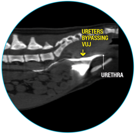
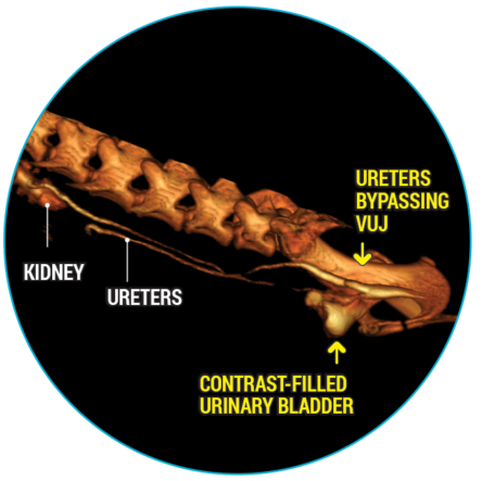
- Bilateral ureteral ectopia with mild right hydroureter
- Possible anterograde urethral reflux/urinary bladder sphincter incompetence
- Intramural bypasses of the vesicourethral junction (VUJ) are considered most likely
How CT Helped in This Case
- Discovered the reason for the patient’s incontinence.
- Provided valuable information for the best treatment plan.
- Allowed complete evaluation of the abdomen in one procedure.
As shown in this case, abdominal CT scanning is an excellent diagnostic imaging modality when further evaluation of the upper urinary tract is needed. A CT scan of the abdomen will also allow for the evaluation of all the intra-abdominal organs (i.e. liver, spleen, stomach, gastrointestinal tract, lymph nodes, reproductive tract, adrenal glands, etc.) as well as the spine and surrounding structures. A recent study states that “computed tomographic excretory urography (CTEU) is a reliable imaging technique for diagnosing canine ectopic ureters.”1 Additionally, a 2017 ACVIM conference proceedings stated that “CTEU has become the more accurate imaging choice for diagnosis of ureteral ectopia.”2
_________
1 Secrest, Bugbee, et als (2016) Comparison of transverse computed tomographic excretory urography images and maximum intensity projection images for diagnosing ectopic ureters in dogs.Vet Radiol Ultrasound. 2017 Mar;58(2):163-168 2 Angela J. Marolf, DVM, DACVR (ACVIM Symposium 2017) When to Consider Abdominal Computed Tomography (CT) Over Abdominal Ultrasound?